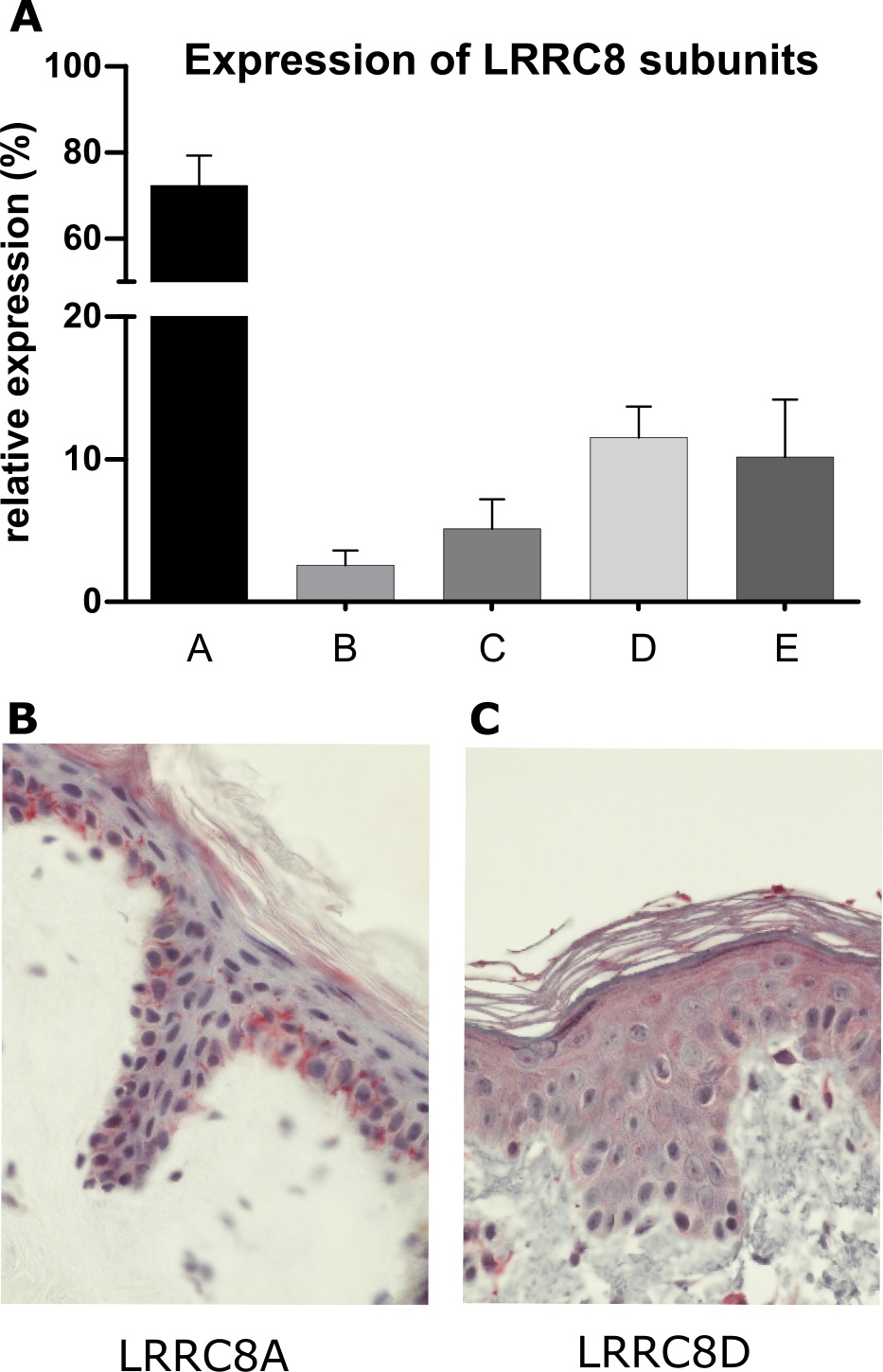
Fig. 4. Expression of LRRC8 in the human epidermis. (A) RNA was isolated from human primary keratinocytes (n=4) and subjected to RNA-Seq. Bars represent transcript levels of LRRC8A-E subunits depicted as relative abundance in relation to all subunits. Human keratinocytes express all LRRC8 subunits, with the highest abundance of LRRC8A and LRRC8D (see also [38]). (B+C) Punch biopsies were taken from healthy donors (approval 144/12 Clinic of the Goethe-University). The Declaration of Helsinki protocols were followed. Specimen were fixed in 4% PFA, paraffin embedded and 4 μm sections were processed routinely. Immunohistochemical stainings was performed with α-LRRC8A (NBP2-32158 from Novus Biologicals) (B) and LRRC8D (11537-1-AP from Proteintech) (C) antibodies. Histofine Simple Stain AP Multi (Nichirei Bioscience) was used for detection. Nuclei were stained with hematoxylin. Images were acquired by using a Nikon Eclipse slide scanning microscope. LRRC8A is preferentially expressed in the basal layer (B), while LRRC8D is more uniformly distributed throughout all epidermal layers (C).